Temporomandibular joint disorders (commonly called TMJ or TMD) affect millions of people and can disrupt everyday activities like eating, speaking and sleeping. While some cases are temporary and resolve with conservative care, others can become a chronic source of discomfort and reduced jaw function. This page explains how the temporomandibular joint works, what can go wrong, how clinicians evaluate TMJ complaints and what treatment approaches are commonly used today.
Understanding How the Jaw’s Anatomy Enables Movement
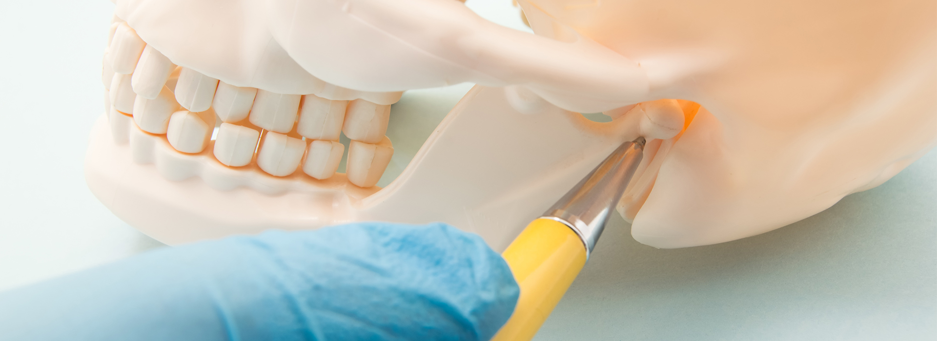
The temporomandibular joint is a paired joint that links the lower jaw (mandible) to the temporal bone of the skull. Each joint contains a small, flexible disc that cushions the rounded condyle of the jaw as it moves inside the joint socket. Because the TMJ allows both hinge-like opening and complex sliding motions, it is uniquely designed to accommodate chewing, speaking and facial expressions.
Muscles, ligaments and tendons around the joint coordinate those motions. When all parts are working smoothly, the jaw opens and closes without pain or restriction. When the disc becomes displaced, muscles go into spasm, or the joint surface becomes inflamed or worn, normal movement can be altered — producing pain, noises or a feeling that the jaw is catching or locking.
Appreciating this anatomy helps explain why symptoms can arise from several different sources: the cartilage-like disc, the bone surfaces, the muscle groups that move the jaw, or the nerves that supply sensation. Successful evaluation and treatment depend on identifying which structures are contributing to a patient’s symptoms.
Common Causes and Who Is More Likely to Develop TMJ Problems
TMJ problems arise from a mix of mechanical, biological and behavioral factors. Repetitive loading of the joint from teeth grinding (bruxism), a direct injury to the jaw, long-standing malocclusion (bite discrepancies), inflammatory conditions such as arthritis, and parafunctional habits like chronic gum chewing can all contribute to joint stress.
Certain risk factors increase the chance that a person will develop symptomatic TMJ issues. Women are diagnosed more often than men, and symptoms most frequently appear during young adulthood and middle age. Psychological stress can heighten muscle tension and the likelihood of clenching or grinding, which in turn aggravates joint structures.
It is also important to remember that not every structural irregularity causes symptoms. Imaging or bite variations may be present without pain. A careful clinical correlation between symptoms and physical findings guides whether and how treatment should proceed.
Recognizing the Key Symptoms That Suggest a TMJ Disorder
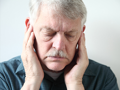
TMJ disorders produce a spectrum of complaints. Jaw pain and tenderness in the face or around the ears are common, as are noises such as clicking, popping or grating with jaw movement. Some patients report episodes where the jaw feels stuck or locked in an open or closed position, or observe restricted range of motion when trying to open widely.
Because the musculature and neural pathways for the jaw overlap with those for the head and neck, TMJ problems can also be associated with headaches, neck and shoulder discomfort, ear-related sensations like fullness or tinnitus, and changes in bite awareness. Symptoms can be intermittent or constant and may fluctuate with stress, sleep quality and oral habits.
Early recognition of patterns — for example, pain that worsens in the morning after nighttime grinding, or progressive limitation of mouth opening — helps clinicians prioritize diagnostic steps and choose the most appropriate interventions.
How TMJ Disorders Are Evaluated in the Clinic
A comprehensive assessment begins with a detailed history and targeted physical exam. Clinicians ask about the onset, duration and character of symptoms and look for signs such as limited opening, joint noises, muscle tenderness and deviations of the jaw during movement. Observing how symptoms change with function helps distinguish muscle-related pain from joint-specific problems.
Imaging is used selectively to clarify joint anatomy or detect degenerative change. Standard radiographs may show bony changes, while advanced imaging such as magnetic resonance imaging (MRI) can reveal disc position and soft-tissue pathology. These tests are ordered based on clinical indications rather than routinely for every patient.
Diagnosis often requires integrating the exam, imaging when appropriate, and an understanding of the patient’s habits and medical history. This integrated approach enables a targeted care plan that addresses the most likely contributors to a patient’s symptoms.
Treatment Strategies: From Self-Care to Clinical Interventions
Initial management of TMJ symptoms typically prioritizes conservative, reversible measures. Patients are commonly encouraged to adopt behavioral changes — such as soft diets, avoiding extreme jaw opening and reducing gum chewing — and to practice stress-reduction techniques that can decrease jaw muscle clenching. Short-term application of cold or moist heat and gentle stretching exercises are often helpful adjuncts.
Oral appliances — such as stabilization splints or night guards — can protect joint surfaces and reduce the effects of nocturnal grinding. These devices are customized by the treating clinician and adjusted over time based on symptom response. Physical therapy approaches that include manual therapy, posture training and home exercises are also valuable for restoring muscle balance and improving function.
When conservative care does not provide adequate relief, additional options may be considered. Targeted injections, occlusal adjustments or coordinated orthodontic or restorative treatments can address specific mechanical contributors in selected cases. Referral to surgical consultation is reserved for persistent, structurally driven problems when less invasive measures have been exhausted.
Long-Term Management and When to Seek Care
Because TMJ disorders can be episodic, establishing a management plan that patients can use at the first sign of recurrence is important. Tracking symptom patterns, maintaining healthy sleep and stress habits, and addressing bruxism proactively reduce the risk of flare-ups. Regular follow-up allows clinicians to modify treatment as needed and monitor for progression.
Anyone experiencing persistent jaw pain, progressive limitation in opening, frequent joint noises with catching or locking, or symptoms that interfere with daily activities should seek a professional evaluation. Early assessment improves the likelihood of identifying reversible contributors and preventing chronicity.
Our team brings an evidence-based, patient-centered approach to evaluating jaw problems. With careful assessment and a stepwise plan that favors conservative care first, we work with patients to restore comfort and function while limiting unnecessary interventions. Inspirational Smiles Orthodontics is committed to coordinating care with other specialists when multidisciplinary management is indicated.
In summary, TMJ disorders encompass a variety of conditions that can affect the joint, muscles and surrounding tissues. Understanding the anatomy, recognizing contributing factors and pursuing a structured evaluation lead to safer, more effective care. If you have questions or would like more information about TMJ assessment and treatment options, please contact us for more information.


